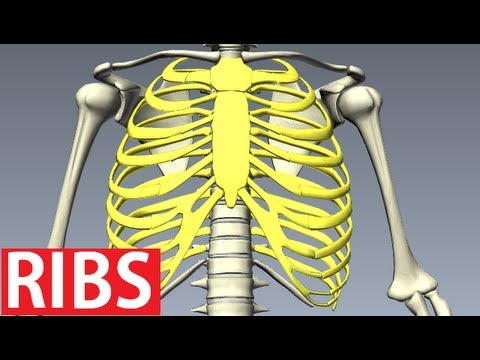
Anatomy Of Rib Cage / The Thoracic Cage Anatomy And Physiology. Of all 24 ribs, the first seven pairs are often labeled as 'true.' these bones are. The costovertebral joint includes a connection between the head of the rib and the inferior costal facet of the vertebral body that the rib is numbered after and a connection between the inferior costal facet of the vertebral body above. However, only seven have a direct articulation with the sternum. Jul 08, 2021 · the ribs are curved, flat bones which form the majority of the thoracic cage. Floating ribs, false ribs, real ribs, manubrium of sternu.
Since our rib cage keeps many of our internal organs safe, there is a tendency to worry when we experience pain in this location. They articulate with the vertebral column posteriorly, and terminate anteriorly as cartilage (known as costal cartilage). Two of the most notable organs behind the left side of the rib cage are the left lung and the spleen. Human ribs diagramnumbered / an inhalation is accomplished when the muscular diaphragm, at the floor of the thoracic cavity, contracts and flattens, while the contraction of intercostal muscles lift the rib cage up and out. The thoracic cage is part of the axial skeleton (also known as the rib cage), and consists of 24 ribs, the sternum, costal cartilage, and the 12 thoracic vertebrae.
Rib Cage Anatomy Bones Of The Thoracic Wall Costae Youtube from i.ytimg.com Rib cage pain can be caused. Of all 24 ribs, the first seven pairs are often labeled as 'true.' these bones are. However, only seven have a direct articulation with the sternum. The thoracic cage consists of the 12 thoracic vertebrae, the associated intervertebral discs, 12 pairs of ribs with their costal cartilages, and the sternum. Elevates the ribs, increasing the thoracic volume. Floating ribs, false ribs, real ribs, manubrium of sternu. The costovertebral joint includes a connection between the head of the rib and the inferior costal facet of the vertebral body that the rib is numbered after and a connection between the inferior costal facet of the vertebral body above. Rib cage anatomy, labeled vector illustration diagram.
Each are symmetrically paired on a right and left side.
The thoracic cage is part of the axial skeleton (also known as the rib cage), and consists of 24 ribs, the sternum, costal cartilage, and the 12 thoracic vertebrae. Rib cage pain can be caused. Related posts of muscle anatomy rib cage muscle anatomy motion. The costovertebral joint includes a connection between the head of the rib and the inferior costal facet of the vertebral body that the rib is numbered after and a connection between the inferior costal facet of the vertebral body above. This furrow isn't present in the 11th and 12th ribs. The sternum is a flat bone that is made up of three parts, the (1) manubrium, (2) body, and the (3) xiphoid process. Anatomy of rib cage area / the thoracic cage anatomy and physiology i : It is made up of 12 pairs of ribs. Sep 27, 2017 · pain under the left rib cage can mean anything from a ruptured spleen. The upper edge is round and the lower sharp. Diagram of human body, liver rib cage, rib cage diagram labeled, rib cage diagram numbered, rib cage diaphragm, rib cage heart, rib cage organs anatomy, rib cage pain, stomach, diagram of human body, liver rib cage, rib cage diagram labeled, rib cage diagram numbered, rib cage diaphragm, rib cage. Anatomy the rib cage is a bony structure found in the chest (thoracic cavity). There are twelve pairs of ribs, all of which articulate with the vertebral column.
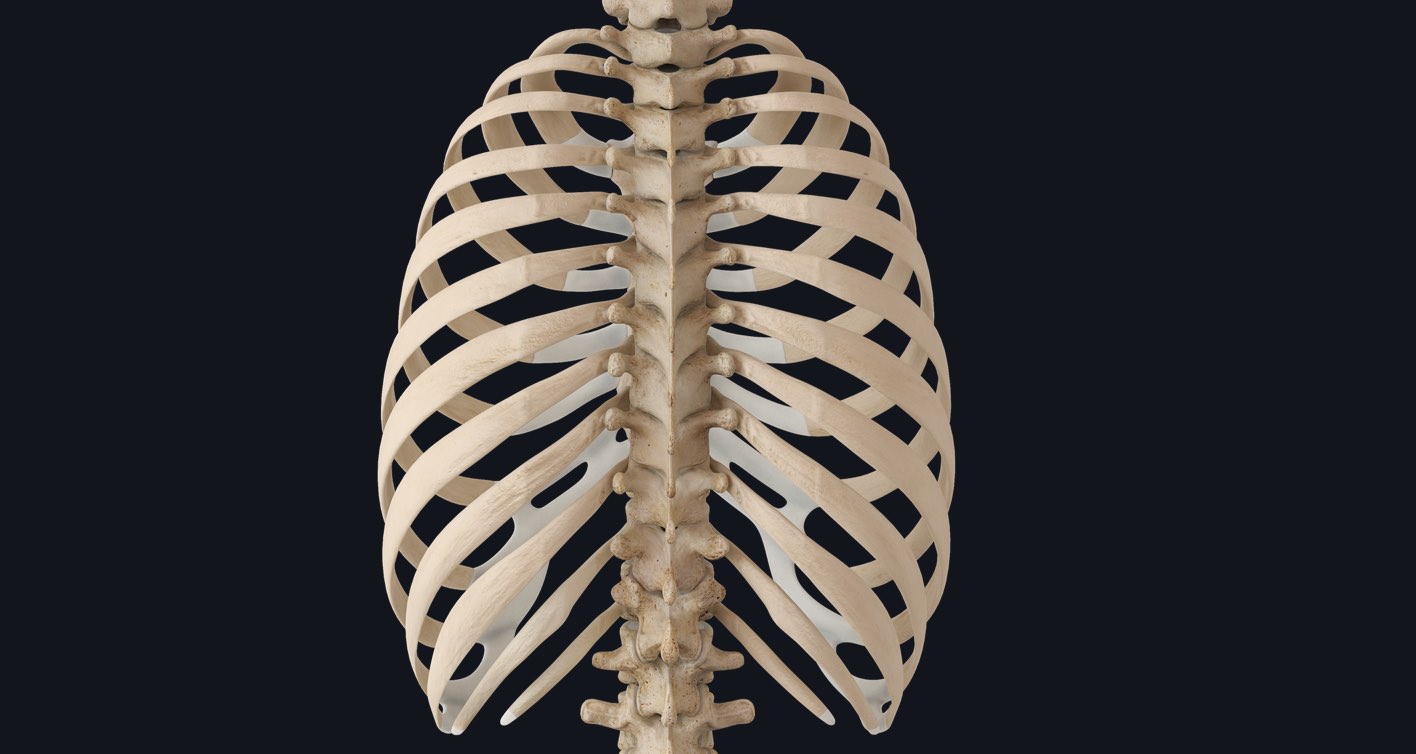
The ribs are curved, flat bones which form the majority of the thoracic cage. The thoracic cage (rib cage) is the skeletal framework of the thoracic wall, which encloses the thoracic cavity. Apr 29, 2019 · the most common causes of rib cage pain are a pulled muscle or bruised ribs. Animated full human body anatomy. There are twelve pairs of ribs, all of which articulate with the vertebral column.
Rib Cage Keeping Things Together In The Thorax Complete Anatomy from cdn.3d4medical.com It is made up of 12 pairs of ribs. They articulate with the vertebral column posteriorly, and terminate anteriorly as cartilage (known as costal cartilage). As part of the bony thorax, the ribs protect the internal thoracic organs. The costovertebral joint includes a connection between the head of the rib and the inferior costal facet of the vertebral body that the rib is numbered after and a connection between the inferior costal facet of the vertebral body above. The upper edge is round and the lower sharp. The bones of the rib cage are the sternum, the 12 thoracic vertebrae and the 12 pairs of ribs. Rib cage anatomy, labeled vector illustration diagram. Each are symmetrically paired on a right and left side.
4 individual objects (spine portion, ribs, cartilages, sternum), sharing the same non overlapping uv layout map, material and pbr textures set.
Floating ribs, false ribs, real ribs, manubrium of sternu. Since our rib cage keeps many of our internal organs safe, there is a tendency to worry when we experience pain in this location. A rib has a flat body, as you can see from the picture of the anatomy of the human rib cage. Apr 29, 2019 · the most common causes of rib cage pain are a pulled muscle or bruised ribs. Related posts of muscle anatomy rib cage muscle anatomy motion. Human ribs diagramnumbered / an inhalation is accomplished when the muscular diaphragm, at the floor of the thoracic cavity, contracts and flattens, while the contraction of intercostal muscles lift the rib cage up and out. The top edge of the manubrium has a depression called the suprasternal or jugular notch. Learn about the anatomy of the human rib cage with this fun educational music video for children and parents. It may occur after an obvious injury or without explanation. The bones of the rib cage are the sternum, the 12 thoracic vertebrae and the 12 pairs of ribs. As part of the bony thorax, the ribs protect the internal thoracic organs. On the interior wall of the rib body is a channel, sulcus costae, with blood vessels and nerves. This furrow isn't present in the 11th and 12th ribs.
The upper edge is round and the lower sharp. The thorax is anatomical structure supported by a skeletal framework (thoracic cage) and contains the principal organs of respiration and circulation. Anatomy of rib cage area / the thoracic cage anatomy and physiology i : The ribs are curved, flat bones which form the majority of the thoracic cage. Rib cage, in vertebrate anatomy, basketlike skeletal structure that forms the chest, or thorax, and is made up of the ribs and their corresponding attachments to the sternum (breastbone) and the vertebral column.
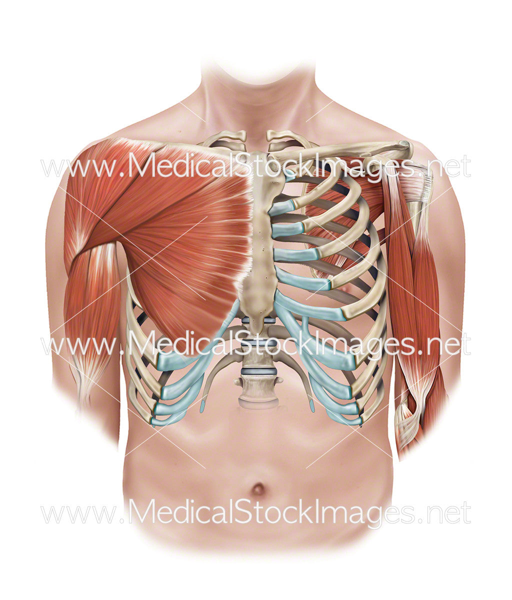
Superficial And Deep Muscles Of The Shoulder And Rib Cage Medical Stock Images Company from cdn.shopify.com The thoracic cage (rib cage) is the skeletal framework of the thoracic wall, which encloses the thoracic cavity. On the interior wall of the rib body is a channel, sulcus costae, with blood vessels and nerves. Human ribs diagramnumbered / an inhalation is accomplished when the muscular diaphragm, at the floor of the thoracic cavity, contracts and flattens, while the contraction of intercostal muscles lift the rib cage up and out. An enlarged or ruptured spleen can cause sudden or chronic pain under the left rib cage that ends up migrating towards the back and/or shoulders. Contributing to their role in protecting the internal thoracic organs. The thoracic cage is part of the axial skeleton (also known as the rib cage), and consists of 24 ribs, the sternum, costal cartilage, and the 12 thoracic vertebrae. There are twelve pairs of ribs, all of which articulate with the vertebral column. The ribs are curved, flat bones which form the majority of the thoracic cage.
The spleen is used to filter red blood cells and hangs in the upper part of the abdomen.
It is made up of 12 pairs of ribs. 16 photos of the rib cage diagram with organs. The rib cage consists of 24 ribs, 12 on either side, and it shields the organs of the chest, including the heart and the lungs, from damage. The thoracic cage (rib cage) is the skeletal framework of the thoracic wall, which encloses the thoracic cavity. This furrow isn't present in the 11th and 12th ribs. Related posts of muscle anatomy rib cage muscle anatomy motion. They articulate with the vertebral column posteriorly, and terminate anteriorly as cartilage (known as costal cartilage). As part of the bony thorax, the ribs protect the internal thoracic organs. Rib cage pain can be caused. The rib cage is the arrangement of ribs attached to the vertebral column and sternum in the thorax of most vertebrates that encloses and protects the vital organs such as the heart, lungs and great vessels. Anatomy of rib cage area / the thoracic cage anatomy and physiology i : The upper edge is round and the lower sharp. Apr 29, 2019 · the most common causes of rib cage pain are a pulled muscle or bruised ribs.
Share :
Post a Comment
for "Anatomy Of Rib Cage / The Thoracic Cage Anatomy And Physiology"

Post a Comment for "Anatomy Of Rib Cage / The Thoracic Cage Anatomy And Physiology"